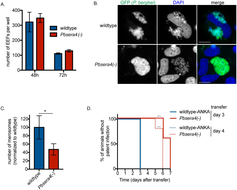
Fig 4. PbSERA4 is not required for liver-stage growth, but is needed for efficient egress from cultured cells.
(A) Huh7 cells were infected with sporozoites of the indicated P. berghei ANKA lines and fixed at the indicated time points. P. berghei exoerythrocytic forms (EEF) were visualized and quantified by fluorescence microscopy. Data is presented as the mean +/- SD. (B) Infected Huh7 cells were fixed and imaged at 65 hours post-infection. At least 50 cells infected with each strain were visualized. More than a quarter of wildtype EEFs were at the advanced stage shown in the upper panel, whereas in the Pbsera4(-)-infected cells, no individual merozoites could be detected in any cell, and none of the Pbsera4(-) parasites had progressed further than the cytomere stage. (C) Merosomes and parasite-containing detached cells were collected from the supernatants of infected Huh7 cells between 65 and 72 hours of liver-stage infection and quantified. Data compiled from five experiments are shown and presented as means +/- SEM. * P<0.05 (ratio paired two-tailed t-test, n = 5 independent experiments). (D) Sub-inoculation from pre-patent Pbsera4(-)-ANKA infected animals. C57BL/6 mice were infected intravenously with 10,000 wildtype-ANKA or Pbsera4(-)-ANKA sporozoites. Blood was transferred from these infected mice to naïve NMRI mice at either 3 or 4 days after infection, and Giemsa-stained blood smears were monitored daily to determine presence of parasites. **, p<0.01, (Log-rank (Mantel-Cox) test, n = 5).

