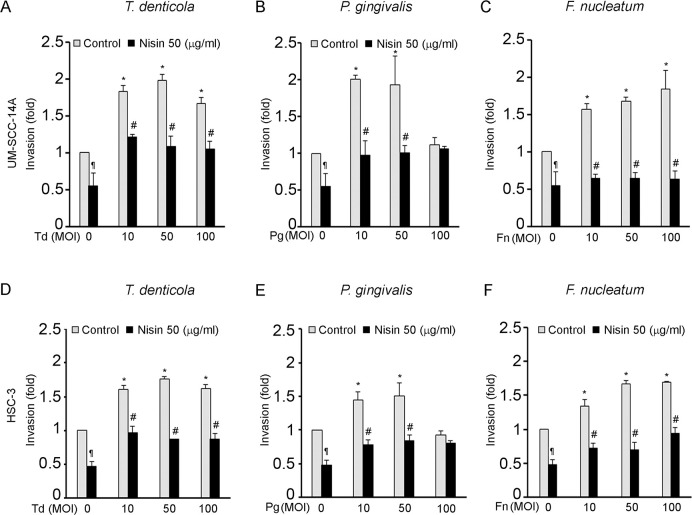
Fig 2. T. denticola, P. gingivalis and F. nucleatum promote invasion of OSCC cells and nisin inhibits that process.
Cells (UM-SCC-14A and HSC-3) were challenged with control medium or media containing different MOIs of T. denticola, P. gingivalis or F. nucleatum for 2 h then treated with nisin (50 μg/ml) for 24 hours and evaluated for invasion potential. Cells that invaded the matrigel-coated membranes were labeled with AM-fluorescent dye and fluorescence intensity was read at 485/520. (A-F) Fold change in invasion relative to the unchallenged cells is illustrated in the graphs. Data represent mean ± SD from three independent experiments. *Comparison between groups relative to their media controls *p≤0.05; #Comparison between groups relative to their matching concentrations of pathogens alone #p≤0.05. ¶Comparison between groups relative to their media control p≤0.05.

