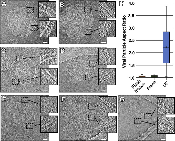
Fig 1. Changes in viral morphology resulting from different purification methods.
Contrast inverted cryo-ET central slices of clarified supernant fluid collected directly from infected Vero cells and either imaged after being flash frozen (A, B), fresh (C, D), or ultracentrifuged (E-G). Inserts represent enlarged regions of the viral glycoprotein layer. (H) Viral particle aspect ratio of all three purification methods (n = 50). Scale bars: (A-G) 50 nm.

