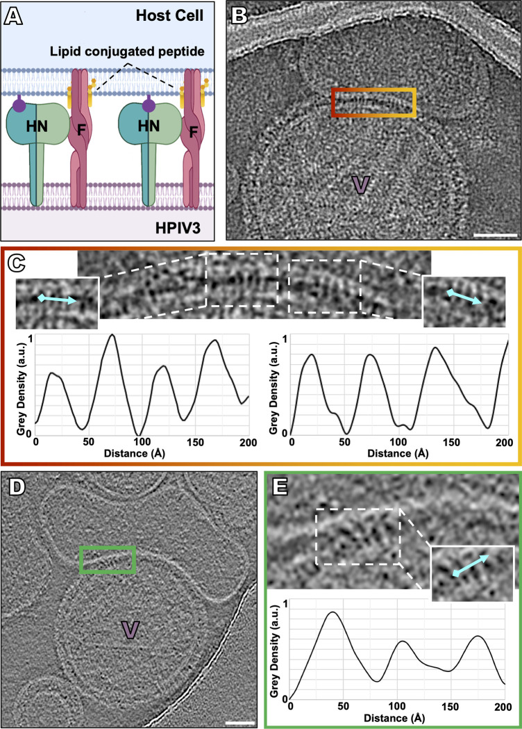
Fig 4. Capture of the transient intermediate state of F with lipid-conjugated fusion inhibitory peptides.
To capture the transient intermediate fusion state just after HN activates F, receptor-bearing target erythrocyte fragment membranes were exposed to virus on grids at 37°C in the presence of lipid conjugated fusion inhibitory peptides (i.e., VIKI-PEG4-chol), prior to vitrification. (A) Schematic of lipid-conjugated peptides inserting into the target cell membrane via their lipid tails and “locking” the extended F in its transient intermediate state, preventing refolding to the post-fusion conformation. (B, D) Contrast-inverted images where viral particles can be observed attached to target erythrocyte fragment membranes using (B) cryo-EM and (D) cryo-ET. (C, E) Enlarged region of interactions between the viral and target erythrocyte fragment membranes where elongated densities linking both membranes are visible. Insets include cyan lines where distance plot measurements were taken. (C,D,E, bottom) Density line plots revealing a repeating 20–35 Å-wide density. Scale bars: (B, E) 50 nm.

