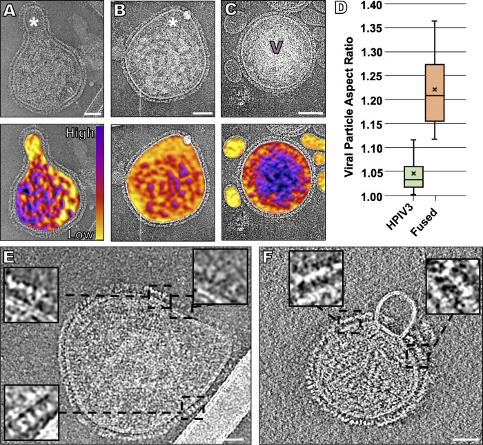
Fig 5. Fusion of HPIV3 with a target erythrocyte fragment membrane.
(A, B) Contrast-inverted viruses that underwent fusion with target erythrocyte fragment membranes (top) with density color representation overlaid below. The purple color represents the dense viral ribonucleoprotein, and the yellow color represents the erythrocyte content. (C) Negative control where grids were kept at 4°C, prior to vitrification to prevent fusion of target erythrocyte membranes with the viruses. (D) Particle aspect ratio of viruses in the presence of zanamivir (also incubated at 4°C, as in C), compared to fused particles (n = 23). (E) Fusion with a small target erythrocyte fragment membrane reveals a lack of prefusion F density near the sites of fusion (inserts). (F) A possible instance of hemifusion where the target erythrocyte fragment membrane surface shows no evidence of surface glycoproteins, and the viral surface shows a lack of prefusion F density near the sites of fusion (inserts). Scale bars: (A-C) and (E, F) 50 nm.

