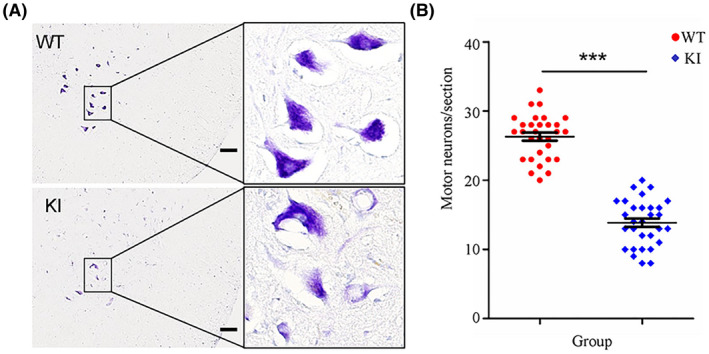
FIGURE 4.

Staining and counting of motor neurons. The lumbar spinal cord tissues of WT and KI rats (n = 3/group) were processed using a standard procedure and sections of lumbar segments at L2 to L4 were stained with cresyl violet. The right ventral horns are shown (A, scale bar = 100 μm). Cells with diameters larger than 20 μm and with a polygonal shape and prominent nucleoli in the ventral horns were counted as motor neurons (B, n = 3 rats/group and 10 serial sections/rat, female). ***P < .001
