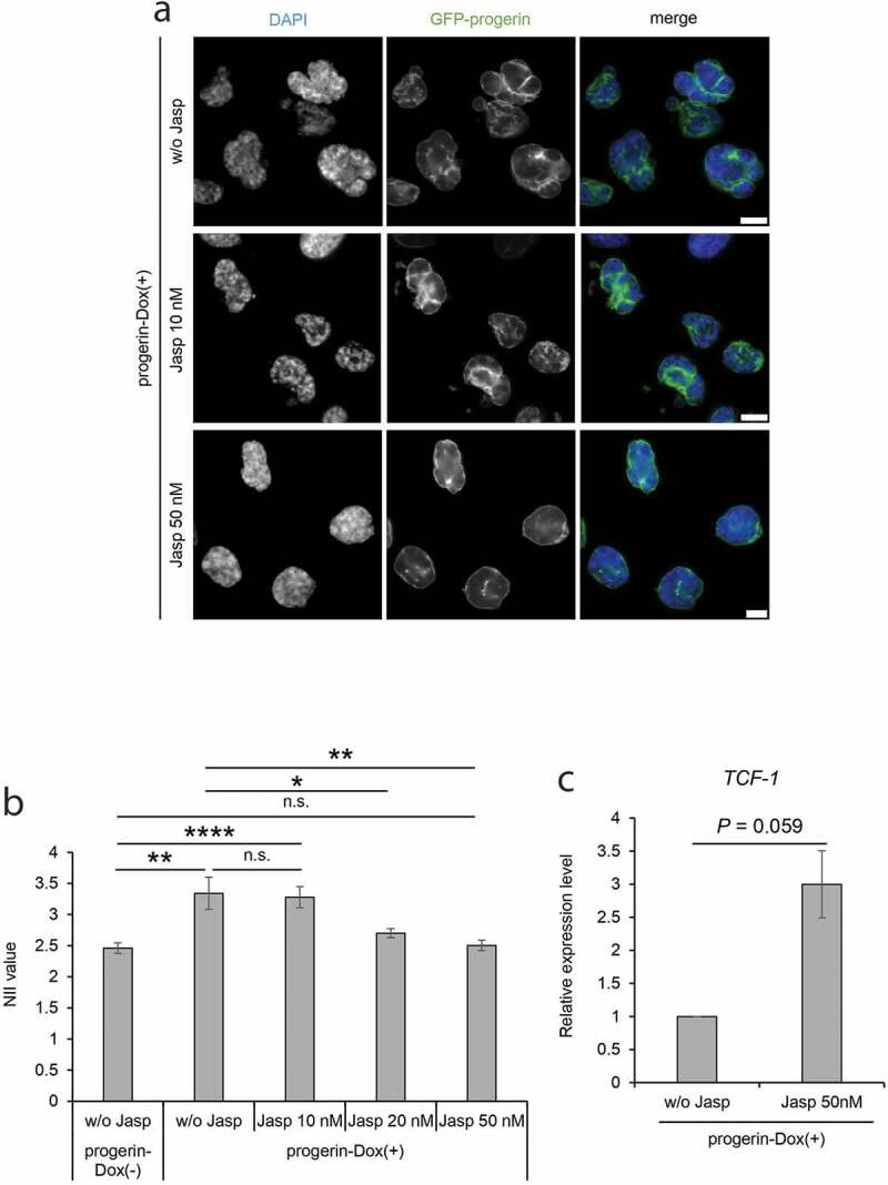
Figure 4.

Complementation of nuclear irregularity of progerin-expressing cells by treatment with jasplakinolide. (a) The progerin-expressing cells were treated with 10 nM or 50 nM jasplakinolide for 24 hours, and then stained with DAPI. DAPI and GFP-progerin images of the cells are compared with those of cells not treated with jasplakinolide (w/o Jasp). Scale bar, 10 μm. (b) The NII values of progerin-Dox(-) cells without jasplakinolide treatment (w/o Jasp) and of progerin-Dox(+) cells with or without jasplakinolide treatment (w/o Jasp, 10 nM, 20 nM or 50 nM) were compared. The NII value was determined in each cell group. Error bars indicate SEM of three independent experiments. For quantification, 526 cells (w/o Jasp, progerin-Dox(-)), 317 cells (w/o Jasp, progerin-Dox(+)), 453 cells (Jasp 10 nM), 630 cells (Jasp 20 nM), and 596 cells (Jasp 50 nM) were analyzed in each experiment. (c) The relative expression level of TCF-1 in progerin-expressing cells treated with jasplakinolide. The expression level was measured by qRT-PCR and was normalized with respect to that of GAPDH gene. The expression level of TCF-1 in progerin-Dox(+) without jasplakinolide treatment was assigned as 1.0. Data shown are mean ± SEM of three independent experiments. n.s., not significant; *, P<0.05; **, P < 0.01; ****, P < 0.0001.
