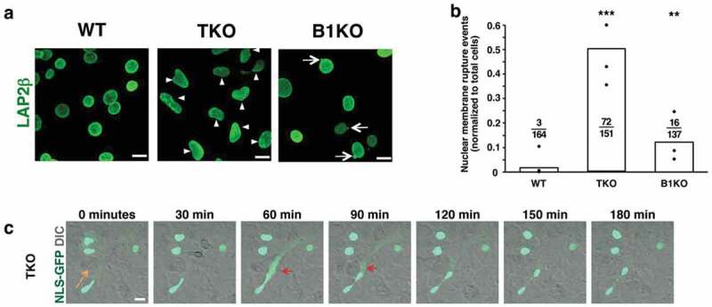
Figure 1.

Abnormal nuclear shape and nuclear membrane ruptures in mouse embryonic fibroblasts (MEFs) lacking all nuclear lamins. (a) Confocal micrographs of Lmna+/+Lmnb1+/+Lmnb2+/+ (wild-type; WT), Lmna–/–Lmnb1–/–Lmnb2–/– (triple knockout; TKO), and Lmna+/+Lmnb1–/–Lmnb2+/+ (Lmnb1 knockout; B1KO) MEFs. Cells are stained with an antibody against LAP2β, an inner nuclear membrane protein (green). Arrowheads point to irregularly shaped nuclei; arrows point to nuclear blebs. Scale bars, 20 μm. (b) Bar graph showing numbers of nuclear membrane rupture events in WT, TKO, and B1KO MEFs. Black circles show the average number of nuclear membrane events (divided by numbers of cells examined) in three independent experiments. Numerical ratios show the total number of nuclear membrane rupture events in all three experiments divided by the total number of cells examined. **P < 0.001; ***P < 0.0001 by χ2 test. (c) Confocal micrographs showing a nuclear membrane rupture (red arrows) in a TKO MEF [expressing a nuclear-localized green fluorescent protein (NLS-GFP)] as it migrated across the field (orange arrow depicts the direction of migration). NLS-GFP is in green; the differential interference contrast (DIC) image is in gray. Scale bar, 20 μm. Reproduced with permission from Chen et al. [16].
