Figure 6.

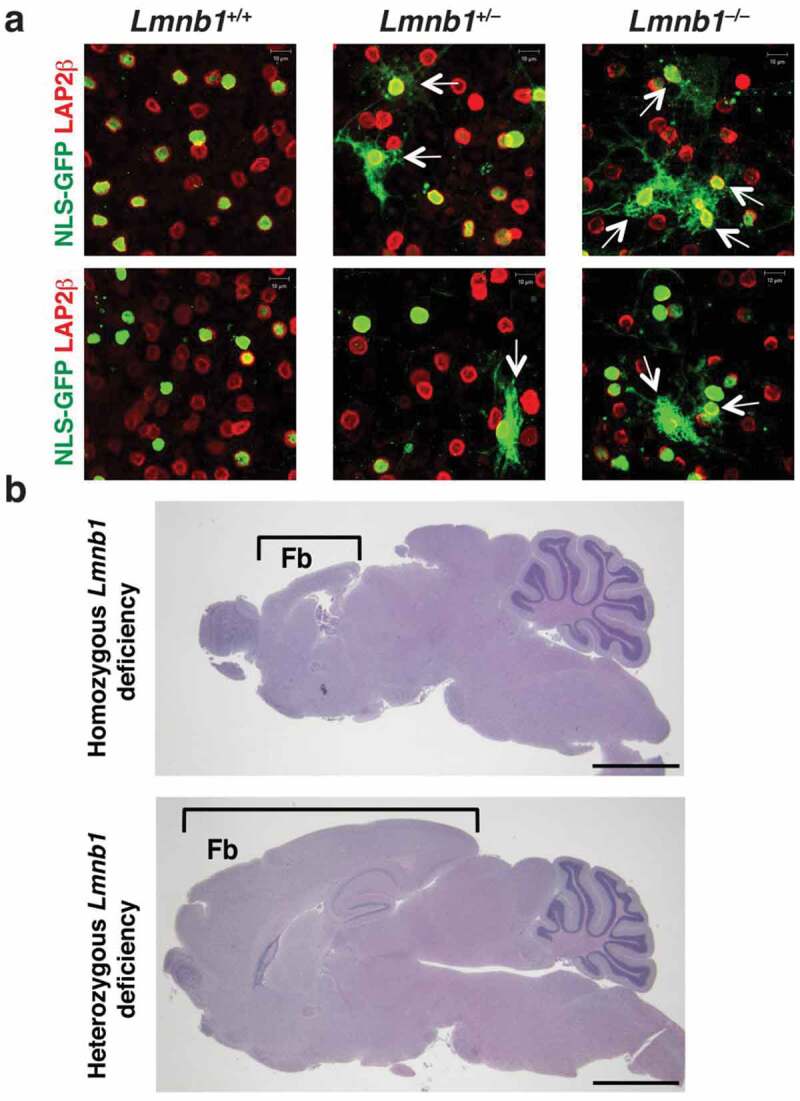
Identifying nuclear membrane ruptures in cultured cells does not always predict pathology in mouse models. (a) Confocal micrographs of NLS-GFP–expressing Lmnb1+/+, Lmnb1+/–, and Lmnb1–/– neurons as they migrate away from cultured neurospheres, revealing nuclear membrane ruptures [escape of NLS-GFP (green) into the cytoplasm] in both Lmnb1+/– and Lmnb1–/– neurons. Ruptures were more frequent in Lmnb1–/ – neurons than in Lmnb1+/– neurons. The neurons were stained with antibodies against LAP2β (red), an inner nuclear membrane protein. White arrows point to cells with nuclear membrane ruptures. Scale bars, 10 μm. (b) Homozygous loss of Lmnb1 in the forebrain in forebrain-specific Lmnb1 knockout mice markedly reduces forebrain size; heterozygous loss of Lmnb1 in the forebrain does not [24]. Brackets indicate the forebrain (Fb). Scale bars, 200 μm. Images in panel b reproduced, with permission, from Coffinier et al. [24].
