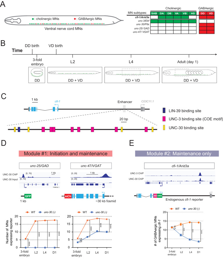
Figure 7. Temporal modularity in UNC-30/Pitx function in GABAergic MNs.
(A) Schematic summarizing the expression of cfi-1/Arid3a, unc-3/Ebf, unc-30/Pitx, unc-25/GAD, and unc-47/VGAT in MN subtypes of the C. elegans ventral nerve cord. (B) Schematic showing time of birth and cell body position of GABAergic nerve cord MNs. DD neurons are born embryonically. VD neurons are born post-embryonically. (C) Bioinformatic analyses predict 4 UNC-30 binding sites (yellow boxes) in the cfi-1 enhancer. The location of UNC-3 and LIN-39 binding sites are also shown. (D) Top: snapshots of UNC-30 ChIP-Seq and input (negative control) signals at the cis-regulatory regions of 2 GABAergic terminal identity genes (unc-25/GAD, unc-47/VGAT). Bottom: quantification of the expression of transgenic reporters in WT and unc-30 (e191) animals at four different developmental stages – 3-fold embryo, L2, L4, and day 1 adults (N = 15). UNC-30 is required for both initiation and maintenance of unc-25/GAD and unc-47/VGAT. ***p<0.001. (E) Top: a snapshot of UNC-3 ChIP-Seq and UNC-30 ChIP-Seq signals at the cfi-1 locus. Bottom: quantification of the number of MNs expressing the endogenous reporter mNG::AID::cfi-1 in WT and unc-30 (e191) animals. All cfi-1-expressing MNs in the ventral cord (cholinergic and GABAergic MNs) were counted in 3-fold embryos due to a lack of a specific marker that labels GABAergic MNs in embryos. Expression of cfi-1 specifically in GABA neurons was quantified at L2, L4, and day 1 adult stages (N ⩾ 12). At those stages, cholinergic MNs were identified based on a fluorescent marker (cho-1::mChOpti), which are ruled out during scoring. GABAergic MNs were scored positive for cfi-1 expression when the mNG::AID::cfi-1 (green) expression co-localized with ttr-39::mCherry (red). N.S.: not significant, ***p<0.001.

