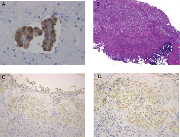
Figure 2.

(A) On pleural effusion cytology, aggregated tumor cells were seen, and were immunocytochemically stained with calretinin (magnification x400). (B) Pleural biopsy revealed that tumor cells invaded the parietal pleura involving the adipose tissue. (Hematoxylin‐eosin staining, magnification x40). (C, D) Immunostaining of tumor nests showed positive staining for IL‐5 (magnification x100 and x200 in (C) and (D), respectively).
