Fig. 6.
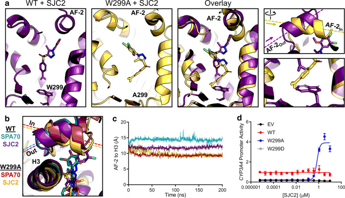
MD predicts an antagonist-to-agonist switch. a Molecular dynamics simulations were performed for WT and W299A PXR LBD in the presence of SJC2. Individual and superimposed images of the ligand-binding pocket are shown for SJC2-bound WT (violet) and W299A (yellow) PXR LBD; the right panels focus on the AF-2 helix and W/A299. b WT or W299A simulations with SJC2 and SPA70 are overlaid. The AF-2 is in the “Out” position for WT simulations and in the “In” position for W299A simulations. c Quantification of the “In” vs. “Out” AF-2 orientations between simulations. The distance from the AF-2 residue F429 to the Helix 3 residue F251 was measured for the duration of each simulation (using α-carbon atoms for measurements). d HepG2 cells were co-transfected with empty vector (EV), WT, W299A, or W299D PXR and a plasmid encoding firefly luciferase under the control of a PXR-responsive CYP3A4 promoter. Cells were treated with SJC2 for 24 h and assayed for luciferase activity

