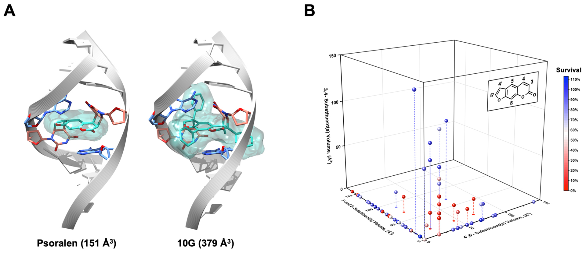
Figure 3.

Size and Positional Constraints on Effective Psoralen Substituents. (a) Using PDB 204D, models of psoralen and 10G ICLs were built to illustrate the possibility for steric clashes with the dsDNA helix when various modifications are made to psoralen. (b) To stratify derivatives according to the sites of their substituents, the psoralen scaffold was divided in three sections: (1) 4′,5′‐ substituents, (2) 5‐ and 8‐ substituents and (3) 3,4‐ substituents. The van der Waals volumes for all substituents were computed in MarvinSketch, and all derivatives were graphed in three dimensions based on the magnitude and position of their substituents’ volumes. To provide context, the volume of a methyl substituent was computed as ~17 Å3 and that of a phenyl was ~80 Å3. Points are colored based on their overall cytotoxicity. Droplines are included when necessary to clarify the three‐dimensional position of several points.
