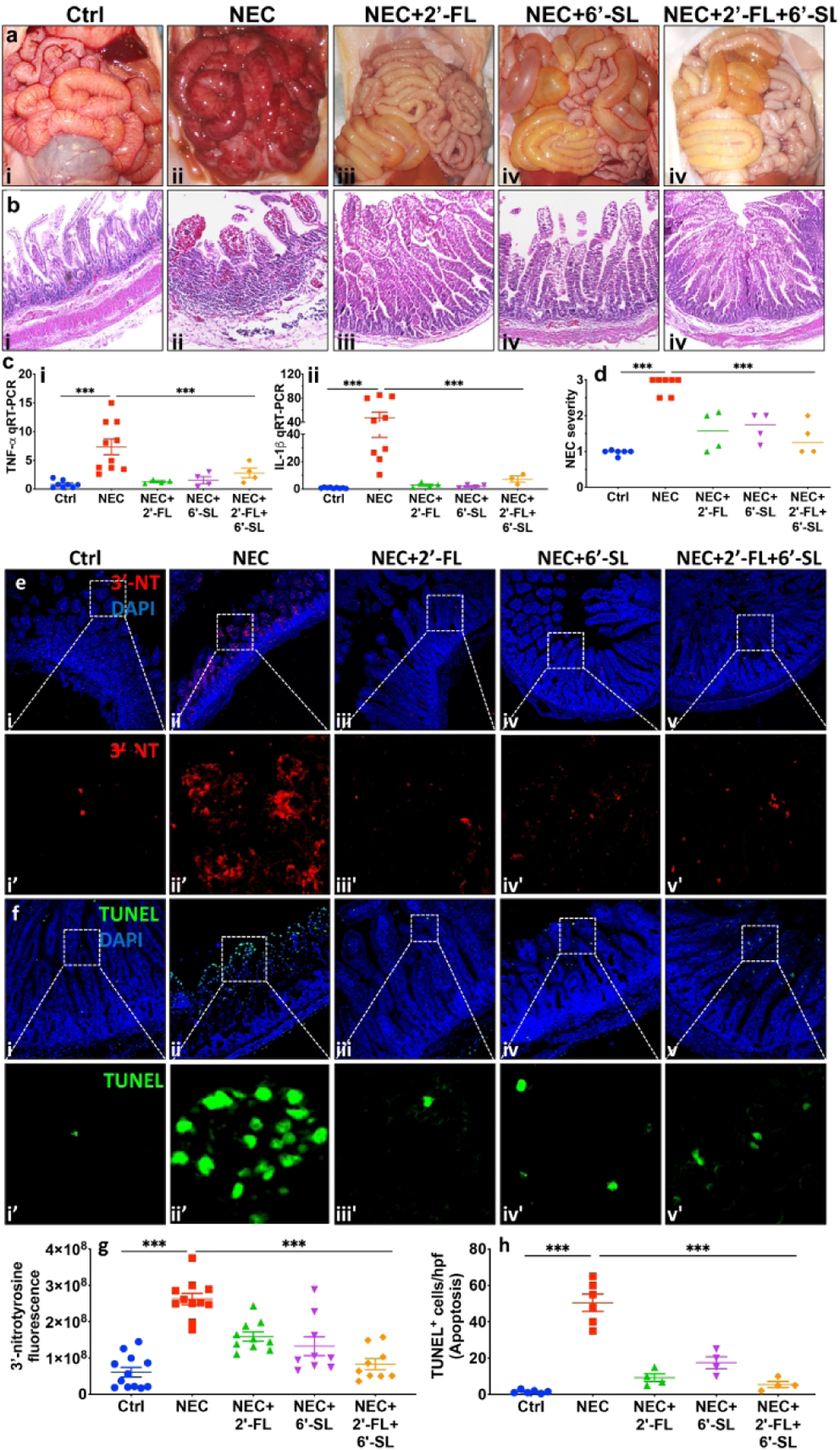
Fig. 3.

Supplementation of nutritional formula with 2’-FL and 6’-SL protects from disease development in a piglet model of necrotizing enterocolitis (NEC). ai-v representative photomicrographs of gross morphology, bi–v Hematoxylin–eosin (H&E)-stained images of premature piglets that were either untreated and euthanized at birth (controls) or induced to develop experimental NEC in the absence or presence of 2’-FL, 6’-SL or 2’-FL+6’-SL, 5μm paraffin imbedded sections of small intestine (ileum), ci-ii quantitative real-time PCR (qRT-PCR) of pro-inflammatory cytokines Tumor necrosis factor-alpha (TNF-α) and Interleukin-beta (IL-1β), d NEC severity scores. *** P<0·001, each dot in dot-graphs represents data from an individual piglet. e-h: 2’-FL) and 6’-SL supplementation prevents small intestinal mucosal damage in a piglet model of necrotizing enterocolitis (NEC). ei- v, i’-v’ immunofluorescence images of 3’-Nitrotryosine (3’-NT, green fluorescence) as an indicator of oxidative mucosal injury, fi- v, i’-v’ immunofluorescence images of TUNEL (green fluorescence) as an indicator of apoptosis injury, in small intestine (ileum) sections of premature piglets subjected to no treatment (Ctrl, control–breast-fed) or experimental NEC treatments without or with supplementation with 2’-FL (10mg/ml/), 6’-SL (10mg/ml), or 2’-FL+6’-SL (5mg/ml, each), g quantification of fluorescent intensity of 3’-NT staining, h quantification of fluorescent intensity of TUNEL staining, measured using ImageJ software, *** P<0·001. Each dot in dot-graphs represents data from an individual piglet.
