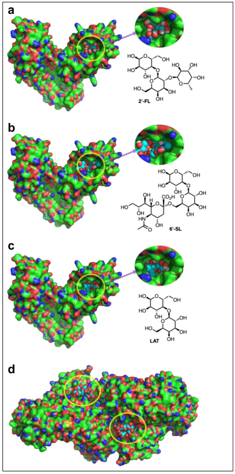
Fig. 6. Docking poses of 2’-FL and 6’-SL, and Lactose (LAT) into the eritoran/LPS binding pocket of the TLR4-MD2 complex.

a CPK model of 2’-FL (cyan carbons) docked to into the x-ray structure of the TLR4-MD2 complex 2Z65, b CPK model of 6’-SL (cyan carbons) docked to into the x-ray structure of the TLR4-MD2 complex 2Z65, c CPK model of Lactose (cyan carbons) docked to into the x-ray structure of the TLR4-MD2 complex 2Z65, d structure of the TLR4-MD2-LPS symmetrical dimer complex 3FXI. The dimerization interface is triggered by LPS (cyan carbons) insertion into the MD2 hydrophobic binding pocket.
