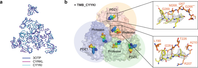
Fig. 5. Co-crystal structures reveal that the tripodal compounds mainly bind to substrate-binding sites of DegP.
a Superposition of three trimers from the previously reported dodecameric structure (PDB code, 3OTP) and our structures with TMB_CYRKL or TMB_CYYKI. b Pentapeptides in DegPS210A•TMB_CYYKI are shown in spheres (rainbow colors from the blue N-terminus to the red C-terminus) on the two substrate-binding sites of DegP. Three DegP monomers are colored differently in surface presentation. Two insets show close-ups of peptapeptides (yellow sticks with electron density maps) and nearby DegP residues (orange sticks) at the PDZ1 pocket (above) and the active-site region (below) of DegP.

