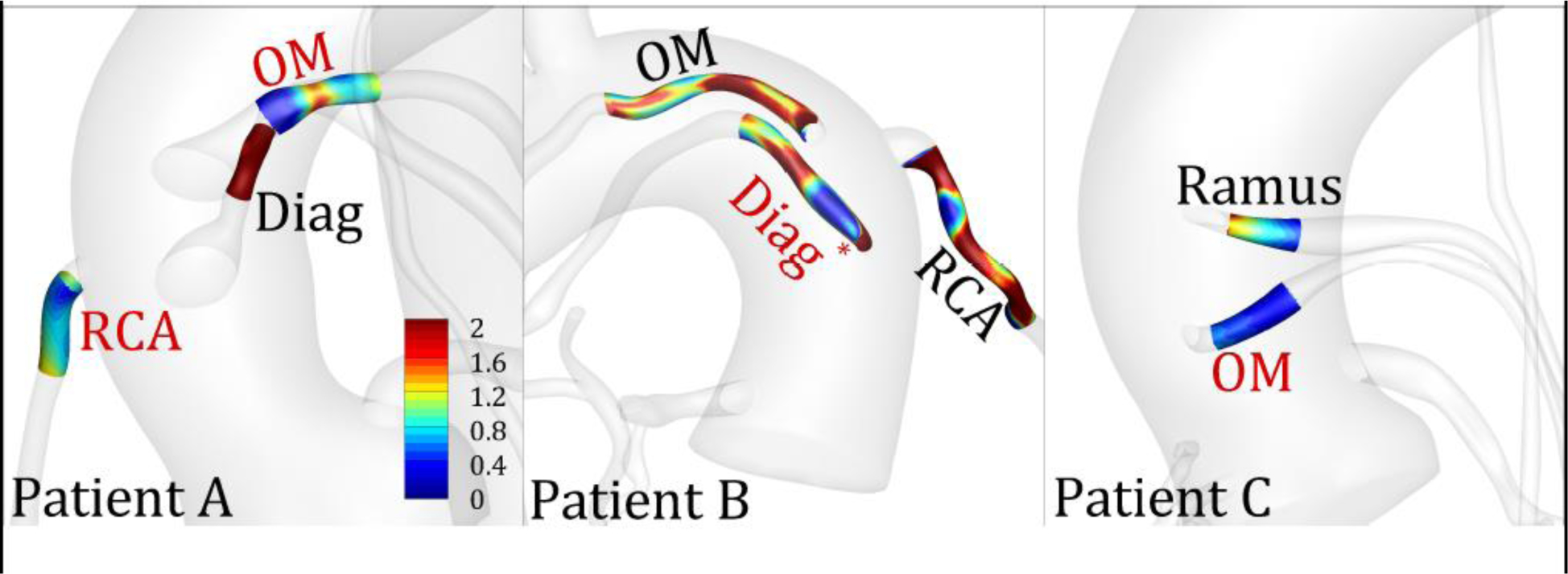
Fig 5:

Distribution of normalized wall shear stress (WSS*) in diseased and control segments in three representative patients. The labels indicate the target vessel to which the SVG was anastomosed. Red and black color fonts represent diseased and control segments, respectively. * indicates that the diseased region was segmented from a stent rather than a stenosis on the CTA. OM, obtuse marginal; Diag, diagonal; RCA, right coronary artery.
