
Figure 1.
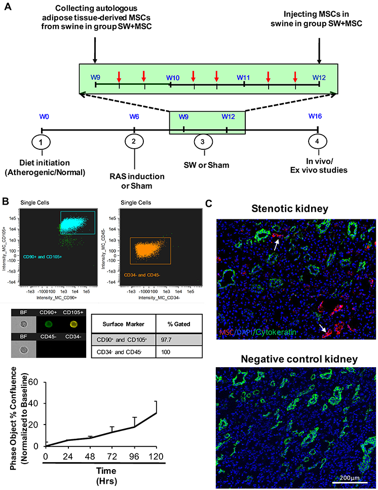
A. Schematic of a 16-wk experimental protocol, with 6 shockwave (SW) therapy sessions delivered over a 3-week regimen (weeks 9, 10, and 11; each session indicated by a red arrow), and autologous adipose tissue-derived mesenchymal stem/stromal cells (MSCs) collection at the beginning of week 9 and injection at the end of week 11 (black arrow). B. Characterization of MSC by markers (CD105 and CD90 by Flow cytometry) and proliferation rate in vitro. Single-color controls were acquired to make a compensation matrix for each test, and the signal visually gauged when gating for positive cells. C. CellTrace™ Far Red-labeled MSCs (white arrow) were detected only in stenotic kidneys injected with MSC (top, x10 images), but not in negative control untreated with MSCs. Cytokeratin (green) was stained as reference for tubular cells.