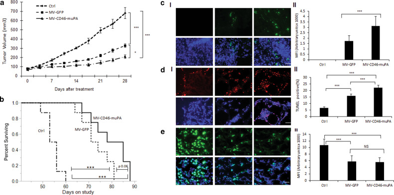
Fig. 4. In vivo effects of MV-CD46-muPA on human colon cancer xenografts.
a Effects of MV-GFP and MV-CD46-muPA on HT-29 tumor progression. Tumor-bearing NSG mice (n = 8 per group) were treated with vehicle (PBS) or MV-GFP and MV-CD46-muPA intravenously, and tumor volume was followed as in methods.. *p < 0.05; **p < 0.01; ***p < 0.001 (days 21, 25, and 28; Tukey–Kramer test). NS: Not significant. b Kaplan–Meier analysis of survival of HT-29 tumor-bearing mice treated with vehicle, MV-GFP, or MV-CD46-muPA. Mice were monitored until they reached killing criteria. *p < 0.05; **p < 0.01. ***p < 0.001 ((log-rank test). NS: Not significant. c Stromal targeting by MV-m-uPA. HT-29 tumor-bearing NSG mice were treated with either vehicle, MV-GFP and MV-CD46-muPA. Immunofluorescence staining for MV-N (green) in tumor tissues obtained at day 3 after virus administration (c, I). Scale bar = 20 μm. d, e. Representative pictures of TUNEL (d, I), and Ki67 (e, I) antibody staining of treated and untreated tumors from mice at day 6 after treatment. Scale bar = 20 μm. Analysis of MV-N (c, II), TUNEL–positive nuclei (d, II), and Ki67 (e, II) staining in vehicle, MV-GFP and MV-CD46-muPA-treated tumors. Bars represent averages±SEM of triplicate experiments (n = 3 per group). *p < 0.05; **p < 0.01; ***p < 0.001 (days 21, 25, and 28; Tukey–Kramer test).

