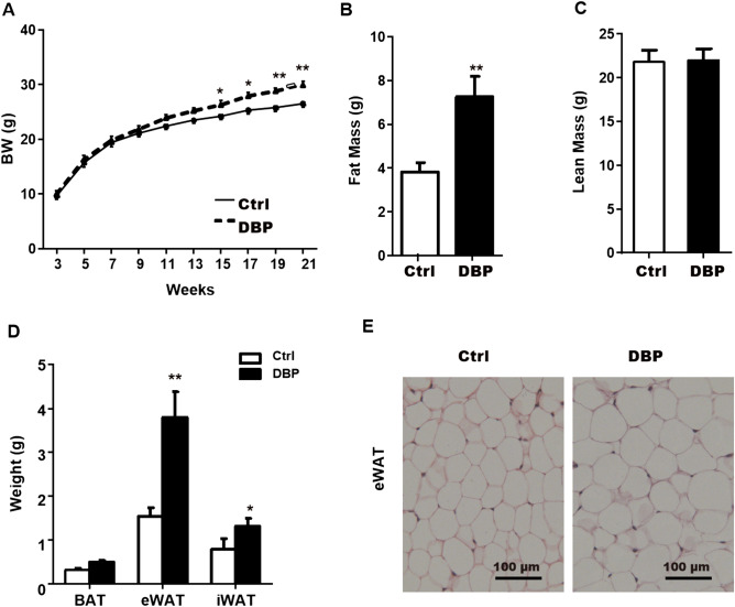
Figure 1.
Effect of in utero DBP exposure on body weight and adipose tissue in offspring. (A) Body weight (BW) during 3–21 weeks in offspring. (B) Fat mass at the 21 week. (C) Lean mass at the 21 week. (D) Adipose tissue weight from different depots. (E) WAT from Ctrl and DBP mice stained with hematoxylin–eosin (H&E), Scale bars = 100 μm. Data are expressed as the mean ± SD, n = 10 per group, *p < 0.05, **p < 0.01 versus the Ctrl group. BW body weight, Ctrl control group, DBP DBP group, WAT White adipose tissue, BAT brown adipose tissue, eWAT epididymal WAT, iWAT inguinal WAT, H&E hematoxylin and eosin.

