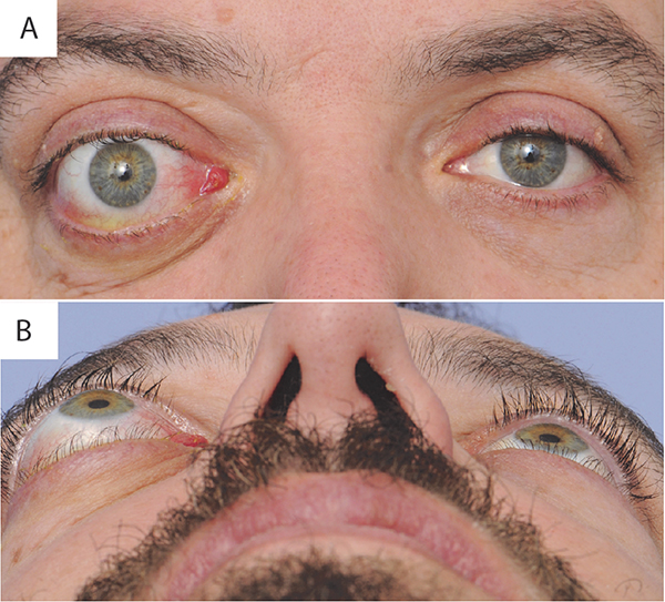
Figure 1:
Thyroid associated ophthalmopathy. A) Portrait view demonstrates axial anterior displacement of the right globe (proptosis), conjunctival injection most prominent along the rectus muscle insertions, lower eyelid retraction, upper eyelid temporal flare, and caruncular edema. B) Worm’s eye view demonstrating the degree of right proptosis relative to the left eye. Note: patient permission was obtained.

