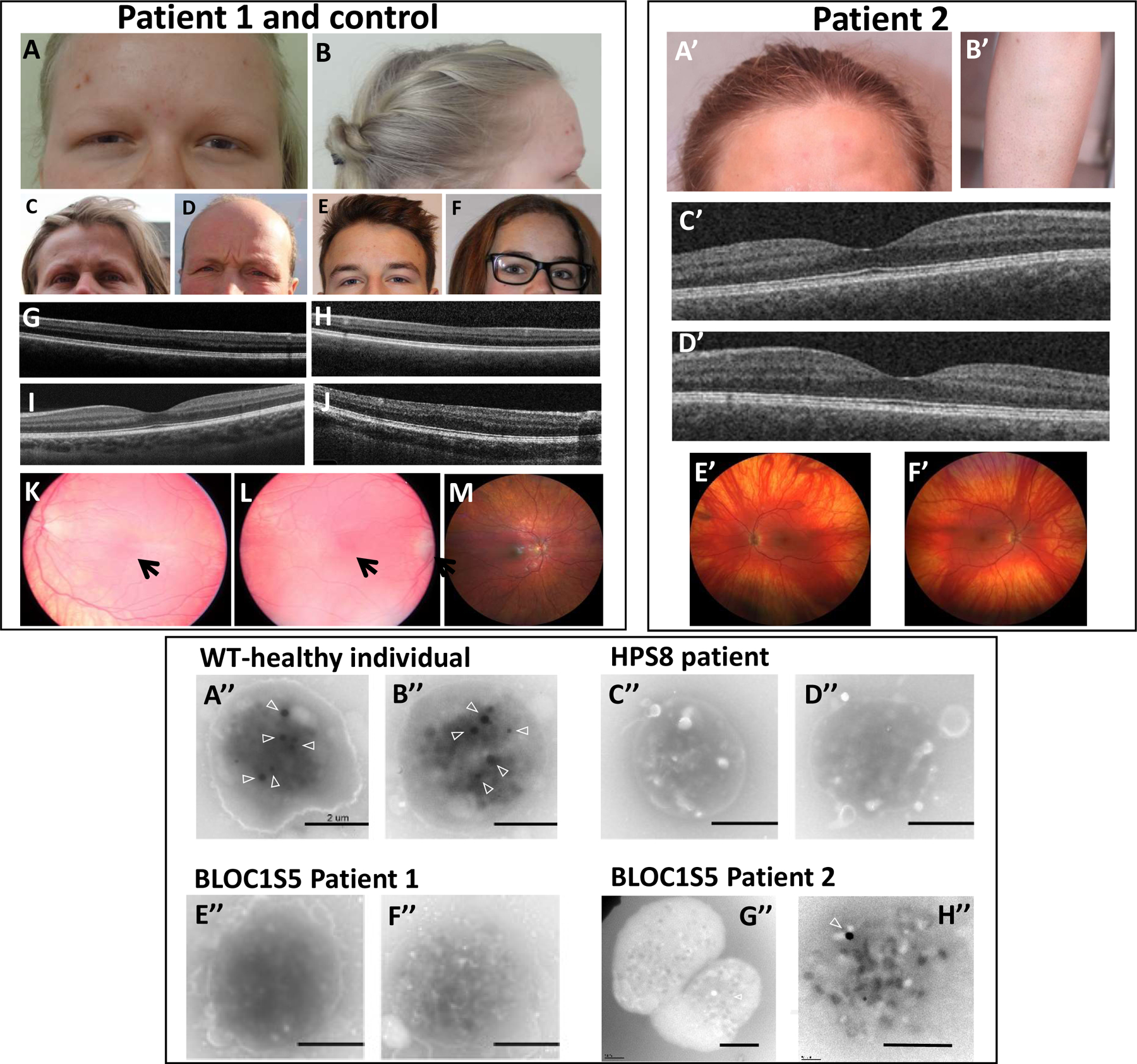
Figure 2. Clinical and platelet phenotype of Patients 1 and 2.

Upper left and right panel: cutaneous and ocular phenotype. Photographs show hypopigmentation of hair and skin of Patient 1 (A,B), compared to her unaffected mother (C), father (D), brother (E) and sister (F), and of Patient 2 (A’,B’). Bilateral foveal hypoplasia is seen in Patient 1 (G: left eye; H: right eye), compared to a normal control (I) and a positive control (OCA1 patient) (J). Patient 2 does not have foveal hypoplasia (C’: left eye; D’: right eye). Retinal hypopigmentation is seen in Patient 1 (K: left eye; L: right eye) compared to a normal control (M). Arrows point to hypoplasia of the macula. Patient 2 has low grade retinal hypopigmentation (E’: left eye; F’: right eye).
Lower panel: whole mount electron microscopy of platelets. Platelets in healthy individuals displayed dense granules typically observed as round electron-dense structures (arrowheads) (A’’-B’’). Platelets from a HPS8 patient were used as a positive control for the absence of platelet dense granules (C’’-D’’). Platelets from the BLOC1S5 Patient 1 harbored no round electron-dense structures (E’’-F’’). Most platelets from the BLOC1S5 Patient 2 harbored no round electron-dense structures (G’’-H’’). Scale bar: 2 μm.
