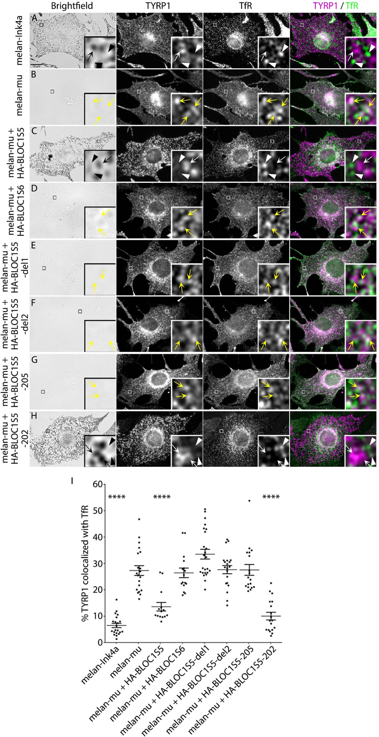
Figure 5. Patient 1-derived BLOC1S5 transgenes do not restore BLOC-1-dependent cargo transport in mouse Bloc1s5mu/mu melanocytes.

(a-h) Mouse melanocyte cell lines melan-Ink4a, melan-mu or melan-mu stably expressing the indicated HA.11-tagged Muted or Pallidin transgenes were fixed, immunolabeled for TYRP1 (magenta) and transferrin receptor (TfR, green), and analyzed by dIFM and by bright field microscopy to detect melanin. Scale, 10 μm. Insets of boxed regions are magnified ten times. TYRP1 localized on TfR-positive compartments (yellow arrowheads) or TYRP1 (white arrow) and TfR (white arrowhead) localized on discrete compartments are indicated. (i) Percent area of overlap between TYRP1 and TfR in melanocyte cell lines. Data are shown as dot plots with mean ± SEM from at least 14 cells representing three independent experiments. Statistical analysis was performed using a one-way ANOVA. ****, P < 0.0001.
