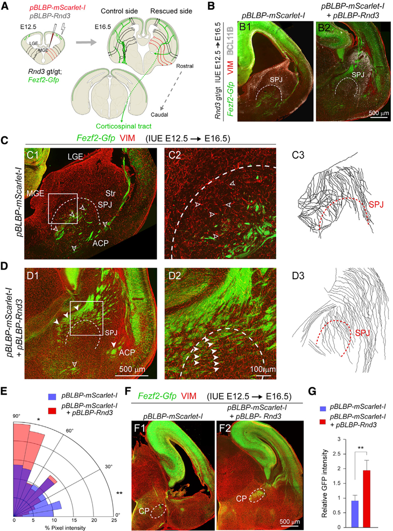
Figure 5. Striatopallidal Pathfinding of CST Axons Depends on RND3 in MGE Radial Glial Cells.

(A) Schematic showing in utero electroporation of pBLBP-Rnd3 and pBLBP-mScarlet-I to the MGE of Rnd3 gt/gt; Fezf2-Gfp brains.
(B) Coronal sections of E16.5 Rnd3 gt/gt; Fezf2-Gfp brains with pBLBP-mScarlet-I (B1) or pBLBP-mScarlet-I+pBLBP-Rnd3 electroporations (B2) at E12.5, immunolabeled for BCL11B (white). The dotted line marks the SPJ boundary. Scale bar: 500 μm.
(C and D) Z-stacked images from E16.5 Rnd3 gt/gt; Fezf2-Gfp brain electroporated with pBLBP-mScarlet-I (C1 and C2) or pBLBP-mScarlet-I-+pBLBP-Rnd3 (D1 and D2), stained for VIM and Fezf2-Gfp, and tracings of the radial glial fibers of the ganglionic eminences (C3 and D3). Scale bars: 500 mm (C1 and D1) and 100 μm (C2 and D2). The open arrowheads show misoriented Fezf2-Gfp+ projections and VIM+ radial fibers (C1 and C2). The solid arrowheads show the rescued Fezf2-Gfp+ and VIM+ radial fibers (D1 and D2).
(E) Polar histogram showing the distribution of orientation of VIM+ radial glial fibers in pBLBP-mScarlet-I (red) or pBLBP-mScarlet-I+pBLBP-Rnd3 (blue) brains of Rnd3 gt/gt; Fezf2-Gfp orthogonal to the VZ of the LGE and MGE in the coronal sections. Pairwise comparison was performed using two-tailed Chi-square test with Yates correction applied (means, *p < 0.05 and **p < 0.01. n = 3/group).
(F) Coronal sections of Rnd3 gt/gt; Fezf2-Gfp brain showing the CST axons at the CP following electroporation with pBLBP-Rnd3+pBLBP-mScarlet-I (F2), or pBLBP-mScarlet-I (F1). Scale bar: 500 μm.
(G) Quantitative analysis of the mean integrated intensity of GFP detected at the CP following pBLBP-Rnd3 in utero electroporation as compared to controls (means ± SEs, n = 3; **p < 0.01; unpaired two-tailed t test).
See also Figure S8.
