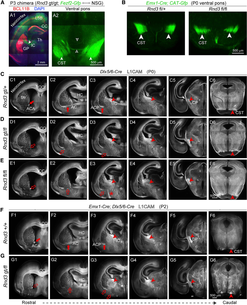
Figure 6. Rnd3 Axon Guidance Phenotypes in the Ventral Forebrain Are Independent of Cortical Fezf2 Expression and Rnd3 Expression in Intermediate Progenitors and Interneurons.

(A) Rnd3 gt/gt; Fezf2-Gfp cells in a wild-type environment, prepared by a blastocyst injection chimera and immunolabeled with antibody against BCL11B (red). A branch of CST showing the decussation defect (open arrow). Scale bars: 2 μm (A1) and 500 μm (A2).
(B) Coronal section of Rnd3 fl/fl; Emx1-Cre mice and Rnd3 fl/+; Emx1-Cre at pons exhibit the size of the CST. Scale bar: 500 μm.
(C–E) L1CAM immunolabeling of Rnd3 fl/fl; Dlx5/6-Cre and Rnd3 gt/fl; Dlx5/6-Cre showing reduced ACA (C1, D1, and E1), reduced and misrouted ACP (D2 and D3, E2 and E3), and stereotypically normal internal capsule (IC), CP, and CST, with deficits highlighted by open arrows and arrowheads and normal features highlighted by solid arrows and arrowheads. Scale bar: 500 μm.
(F and G) Rnd3 gt/fl; Emx1-Cre; Dlx5/6-Cre mice showed a reduction of the ACA and failure to cross the midline of the ACP, and no reduction in CST projections at CP and pons. Scale bar: 500 μm.
See also Figures S4 and S8.
