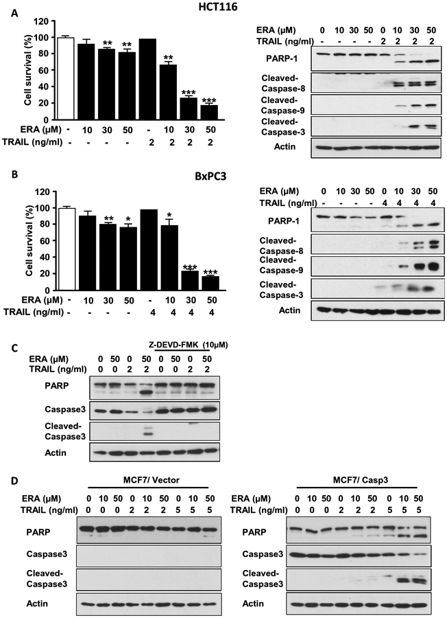
Figure 1. ERA enhanced TRAIL-induced apoptosis by promoting activation of caspases.

Human colorectal carcinoma HCT116 cells (A) and human pancreatic adenocarcinoma BxPC3 cells (B) were pretreated with ERA (10-50 μM) for 20 h and treated with TRAIL (2 ng or 4 ng/ml) for 4 h in the presence of ERA. Cell survival was analyzed using trypan blue exclusion assay (left panels). Western blotting was used to detect the expression of indicated proteins (right panels). (C) HCT116 cells were pretreated with ERA (50 μM) for 19 h and then pretreated with Z-DEVD-FMK (10 μM) for 1 h prior to TRAIL (2 ng/ml) treatment for 4 h in the presence of ERA and Z-DEVD-FMK. Western blotting was used to detect the expression of indicated proteins. (D) MCF7/Vector (left panel) and MCF7/Casp3 (right panel) cells were pretreated with ERA (10 or 50 μM) for 20 h and treated with TRAIL (2 or 5 ng/ml) for 4 h in the presence of ERA. Western blotting was used to detect the expression of indicated proteins. Error bars represent the mean ± SD from triplicate experiments. For statistical analysis, the Student t test (two-sided, paired) was used. P-values: ns, not significant; *P < 0.05, **P < 0.01, ***P < 0.001
