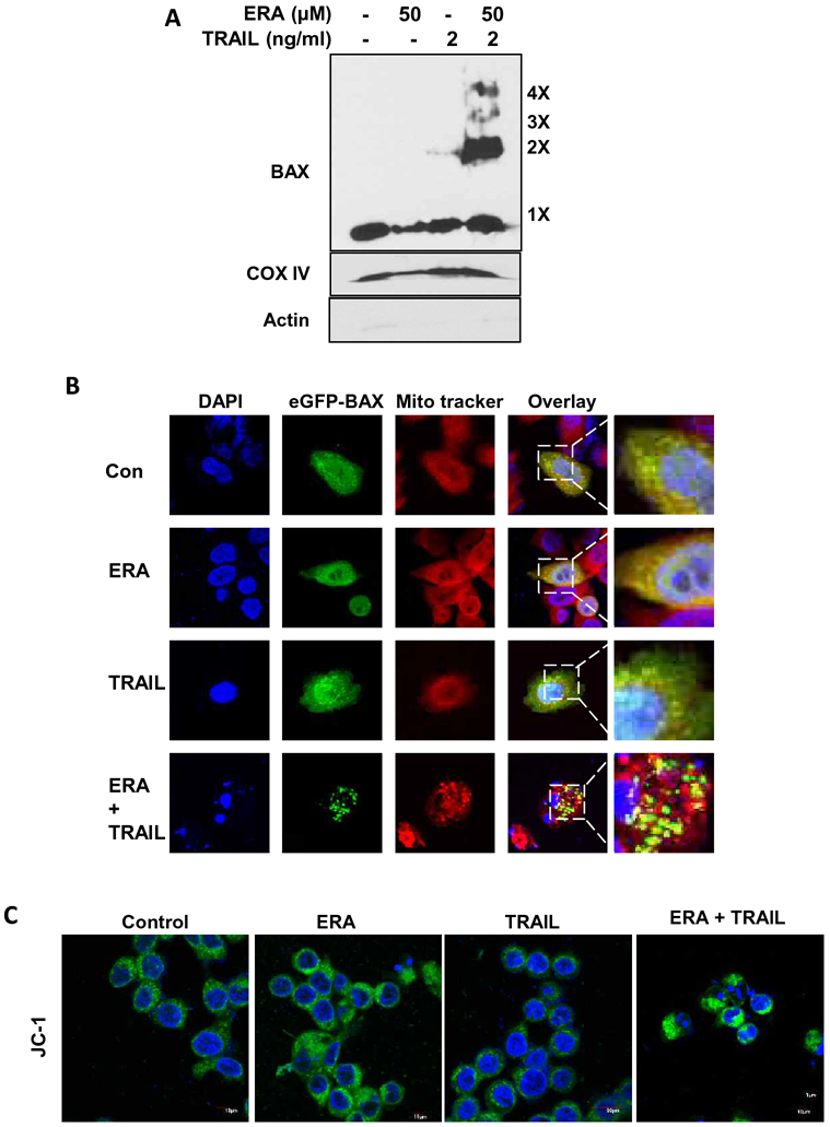
Figure 4. BAX oligomerization and disruption of mitochondrial membrane potential during the combined treatment of ERA and TRAIL in HCT116 cells.

(A) Cells were pretreated with ERA (50 μM) for 20 h and treated with TRAIL (2 ng/ml) for 4 h in the presence of ERA. After treatment, mitochondrial fractions were isolated and cross-linked and then subjected to immunoblotting with an antibody to BAX. BAX monomers (1×) and multimers (2×–4×) are indicated. We used actin (cytosolic marker) and COX IV (mitochondrial marker as fractional markers. (B) Cells were transfected with GFP-BAX plasmids. The cells were pretreated with ERA (50 μM) for 20 h and treated with TRAIL (2 ng/ml) for an additional 4 h. After treatment, the cells were stained with MitoTracker Red. Co-localization of GFP-BAX puncta and MitoTracker Red was examined using a confocal microscope. (C) Cells were pretreated with ERA (50 μM) for 20 h and treated with TRAIL (2 ng/ml) for 4 h in the presence of ERA. After treatment, the cells were stained with JC-1 probe and detected using a confocal microscope. Green fluorescence represents the monomeric form of JC-1, indicating low mitochondrial membrane potential (ΔΨm).
