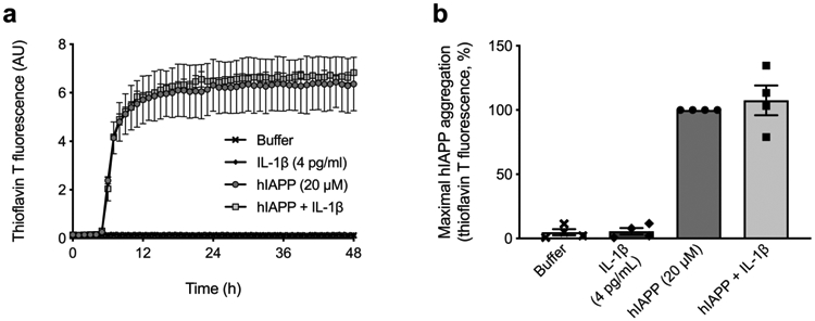
Fig. 3.

Low concentration IL-1β does not increase hIAPP aggregation kinetics. (a) Aggregation of hIAPP was carried out in the absence (circles) or presence (squares) of 4 pg/ml IL-1β and monitored for 48 h by Thioflavin T fluorescence in a cell-free system. One representative experiment is shown with technical replicates. (b) Quantification of maximal hIAPP aggregation revealed no differences in hIAPP aggregation without (black circles) or with (black squares) 4 pg/ml IL-1β. In the absence of hIAPP, neither buffer nor IL-1β increased Thioflavin T signal. Data are presented as mean ± SEM. n=4. AU, arbitrary units
