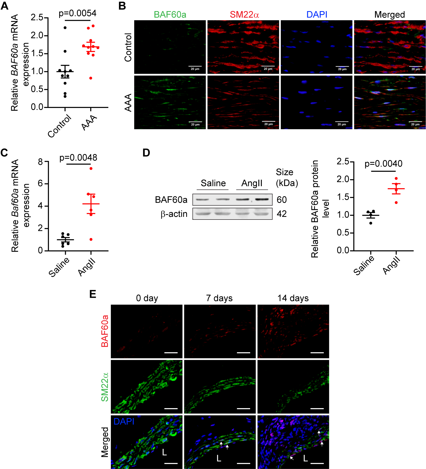
Figure 1. BAF60a is upregulated in human and murine AAA lesions.

A, BAF60a mRNA expression was determined by qPCR in human AAA lesions (n=10) and control abdominal aortas (n=10). B, Representative immunofluorescence staining of BAF60a (green) in human AAA samples (n=3) and control abdominal aortas (n=3). SM22α, red; DAPI blue. Scale bar=20 μm. For the images, the upper side is the adventitial side and lower side is the lumen side. C-D, Relative BAF60a mRNA and protein levels were determined by qPCR (C, n=6 for each group) and Western blotting (D, n=4 for each group) in abdominal aortas of C57BL/6J mice infused with saline or AngII for 28 days. E, Representative immunofluorescence staining of BAF60a (red) in the infrarenal abdominal aortas of C57BL/6J mice at 0, 7, or 14 days after elastase exposure (n=3 for each group). SM22α, green; DAPI, blue. Scale bar=100 μm; L, lumen. Data are presented as mean±SEM. Student’s t-test for A, C and D.
