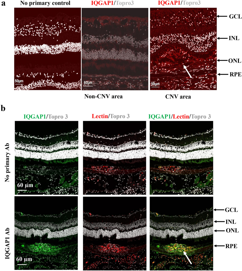
Fig. 2. IQGAP1 is located at CNV lesions.
(a) Immunostaining of IQGAP1 in retinal sections of human donor eyes with AMD (Red: IQGAP1; Gray: Topro 3 to stain nuclei); (b) immunostaining of IQGAP1 and isolectin in retinal cryosections of wild type mice treated with laser (Green: IQGAP1; Red: isolectin; Gray: Topro3 to stain nuclei; GCL, ganglion cell layer; INL, inner nuclear layer; ONL, outer nuclear layer; RPE, retinal pigment epithelium; the white arrows point to type 2 CNV).

