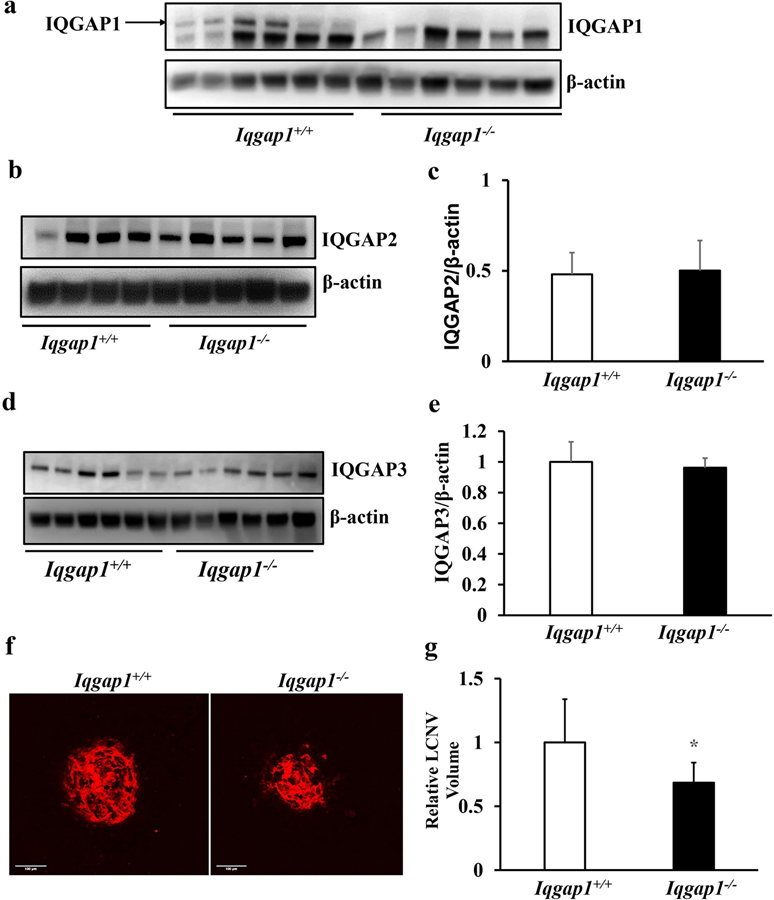
Fig. 3. Iqgap1−/− mice show reduced CNV in a laser-induced CNV model.
Western blots of (a) IQGAP1, (b-c) IQGAP2, and (d-e) IQGAP3 in RPE/choroids of Iqgap1+/+ and Iqgap1−/− mice 7 days post laser treatment (b and d, representative gel images, and c and e, quantification of densitometry); (f) Representative images of RPE/choroid flat mounts and (g) quantification of CNV lesion (*p<0.05 vs. Iqgap1+/+ mice, n= 52 spots from 14 mice) in Iqgap1+/+ and Iqgap1−/− mice 7 days post laser treatment.

