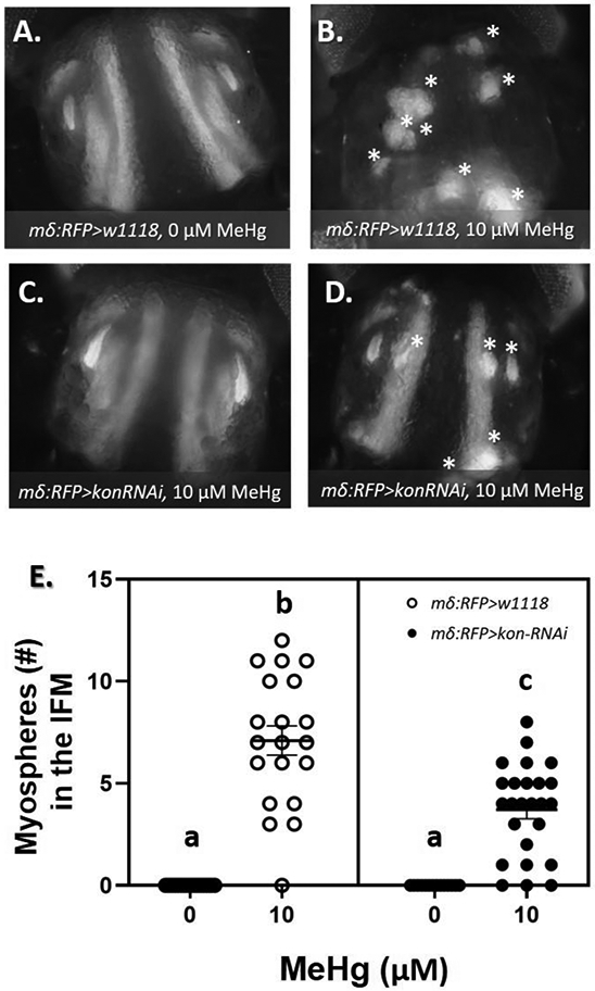
Fig. 7. Frequency of IFM morphological phenotypes after combined MeHg exposure and reduced levels of kon.

(A - D) Representative images of 49 - 57 h APF pupae after IFM-restricted kon knockdown (mδRFP>w1118 or mδRFP>kon-RNAi) and 0 or 10 μM larval MeHg exposure. At least 20 images were taken per group. Asterisks (*) indicate myospheres in the IFM. (E) Quantification of the average ± SD number of myospheres in the IFM reveal a partial rescue of the muscle attachment phenotype induced by developmental exposure to 10 μM MeHg. Letters indicate pair-wise significant differences where p < 0.001 (Two-way ANOVA with Tukey’s HSD, n > 20 pupae per group).
