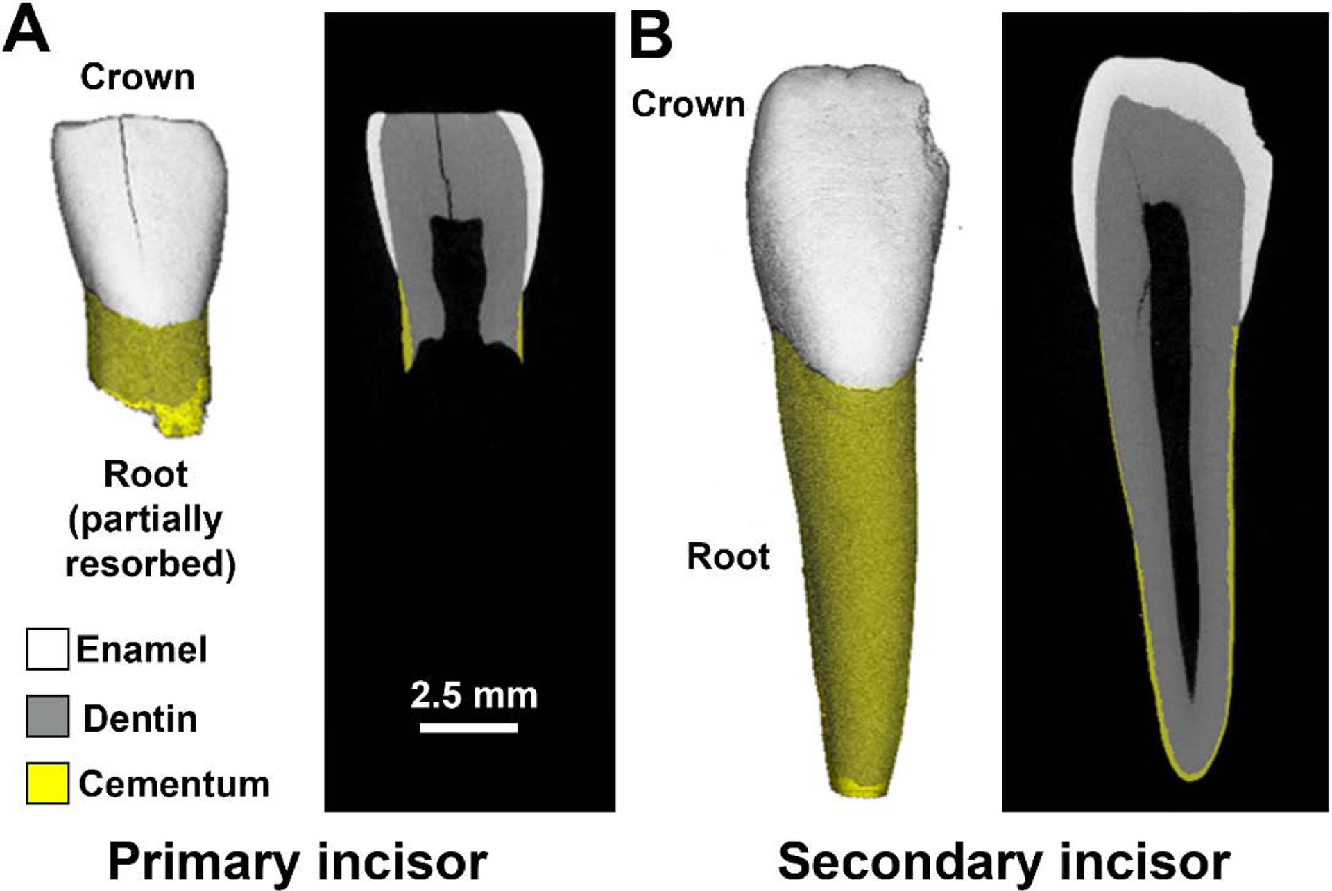
Fig. 1.

Dental Mineralized Tissues in Healthy Teeth. A) Human primary incisor shown by micro-CT in 3D and 2D exhibiting enamel (white), dentin (gray), and cementum (yellow). Note the root has undergone partial resorption as part of the physiological process of exfoliation. B) Human secondary incisor shown by micro-CT in 3D and 2D exhibiting the crown and a full-length root.
