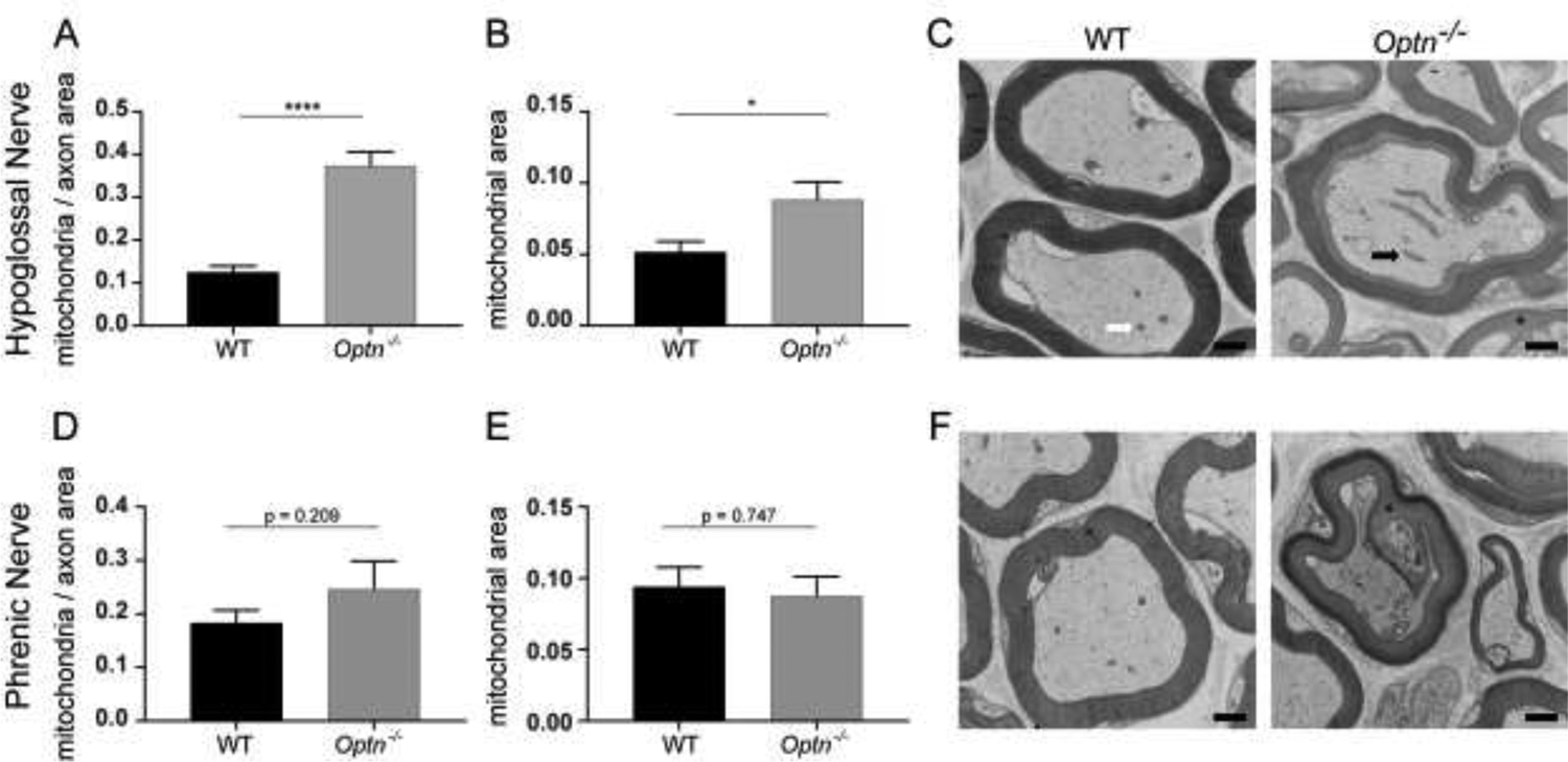
FIGURE 6: Enlarged Mitochondria Accumulate in Respiratory Motor Neuron Axons of Optn−/− Mice.

Panels A and D: Quantification of mitochondria within hypoglossal nerve axons and phrenic nerve axons, respectively, normalized to axonal area. ****p<0.0001. Panels B and E: Quantification of axonal mitochondrial size within the hypoglossal nerve axons and phrenic nerve axons, respectively. Panels C and F: Representative electron micrographs of hypoglossal nerve axons and phrenic nerve axons. The white arrow points to a representative healthy mitochondrion and the black arrow points to a representative enlarged mitochondrion. The blask asterisk identifies points of dysmyelination. Scale bar = 500nm.
