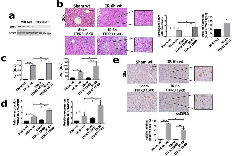
Figure 6 – Hepatic ischemia-reperfusion injury is worse in ITPR3 LSKO mice.
(a) Western blot of total liver protein of ITPR3 in wild-type (WT) and liver-specific-knockout mice for ITPR3 (ITPR3 LSKO). (b) Photomicrograph of liver showing an increase in necrotic areas in LSKO relative to WT.Serum levels of (c) AST and ALT, relative mRNA levels for (d) IL-6 and IL-1. (e) Immunohistochemistry for ssDNA (in brown) and its quantification showing extent of apoptosis is attenuated in ITPR3 LSKO and WT mice. Scale bar, 50 μm. Values expressed as mean ± SEM, n=5, *p<0.05 **p<0.01 ***p<0.001.

