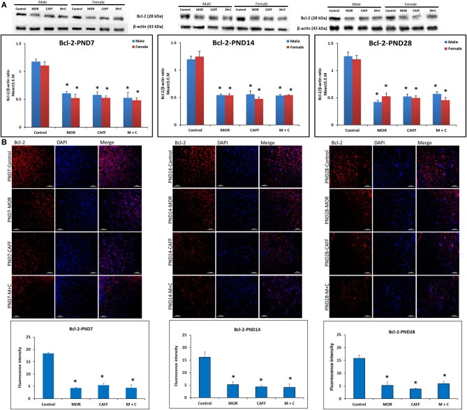
Figure 2.
Expression of anti-apoptosis marker Bcl-2 in rat pups brain. (A) Western blots and densitometry graphs, (B) immunofluorescence images in Control, MOR, CAFF, and M+C treated pups brain tissues at PND 7, PND 14, and PND 28. (A) All blots are representative of four different experiments with similar results. β-Actin was used as a loading control. (B) Representative Immunofluorescence microscopy images and fluorescence intensity graphs of Bcl-2 markers. MOR, CAFF, and M+C groups marked increase in the expression of the anti-apoptosis marker Bcl-2 (red) compared to control. Nuclei were stained with DAPI (blue). Bar scale = 100 μm. Values are expressed as mean ± S.E.M. *P < 0.01 vs. Control.

