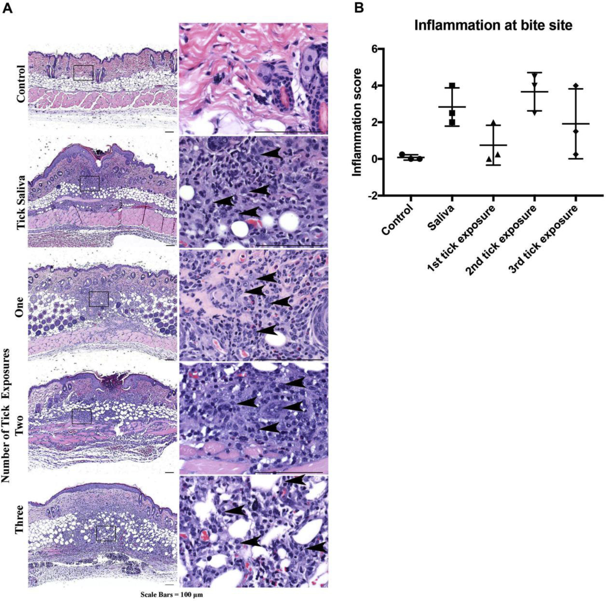
Figure 6. Mouse histopathology of normal non-bite and bite site skin after repeat tick exposures or saliva immunization.

A Compared to non-bite, skin at the bite sites (harvested 4 days after tick attachment) show increased inflammation composed predominantly of mononuclear cells (arrowheads) over neutrophils/eosinophils (representative images). B Inflammation severity scores. Data are represented as mean ± SEM.
