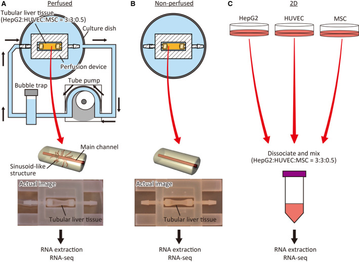
Fig. 1.

Schematic representation of the experimental design. (A) In the perfused group, the tubular liver tissue was cultured under perfusion with medium, using a tube pump. Total RNA was extracted from tissue detached from the device and used for RNA‐seq. (B) In the nonperfused group, the tubular liver tissue was submerged in the medium and statically cultured. Total RNA was extracted from the tissue detached from the device and subjected to RNA‐seq. (C) In the 2D‐cultured group, hepatocellular carcinoma cell line HepG2, HUVECs, and MSCs were cultured in cell culture dishes. The cells were dissociated, mixed at the same ratio as tubular liver tissues, and used for RNA‐seq after total RNA extraction.
