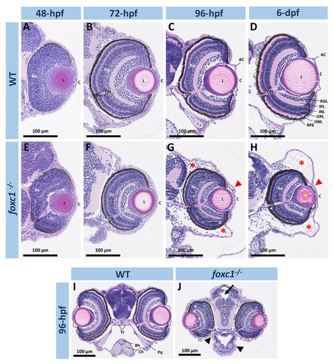
Figure 2.

Histological analysis of ocular and craniofacial anomalies in the foxc1−/−embryos. (A–H) 20× H&E stained histology sections of the eye at 48-, 72-, 96-hpf and 6-dpf of WT and foxc1−/− double knockout embryos. Mutant embryos showed microphthalmia, absence of the anterior chamber (red arrowheads), periocular edema (red asterisks) and lens opacity (yellow asterisk). (I and J) 10× H&E sections of the head at 96-hpf showing smaller head, hydrocephalus (black arrow) and facial cartilage defects (black arrowhead). AC, anterior chamber; C, cornea; L, lens; Re, retina; ON, optic nerve; RGL, retinal ganglion layer; IPL, inner plexiform layer; INL, inner nuclear layer; OPL, outer plexiform layer; ONL, outer nuclear layer; RPE, retinal pigmented epithelium; Bh, basihyal; Ch, ceratohyal; Tr, trabecular; Pq, palatoquadrate.
