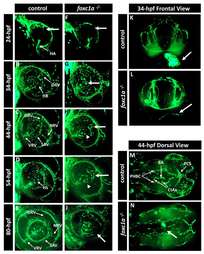
Figure 3.

foxc1a −/− knockout embryos present with defects in the development of the eye vasculature. (A–J) Three-dimensional maximum projection images of the ocular vasculature in live control (A–E) or foxc1a−/− knockout (F–J) embryos carrying the fli1a:EGFP transgene at 24-, 34-, 44-, 54- and 80-hpf. Mutant embryos showed abnormal development of the superficial choroidal vessel (white arrow) and amorphous hyaloid system (white arrowhead). (K and L) Three-dimensional maximum projection images of the head vasculature and heart from the frontal view of 34-hpf control and mutant embryos. Mutant embryos do not show any fli1a:EGFP fluorescence in the heart region (white arrow in L). (M and N) Three-dimensional maximum projection images of the head vasculature from the dorsal view of 44-hpf control and mutant embryos. Mutant embryos show defects in the brain vasculature development such as absence of the central arteries (white arrow in N). HA, hyaloid artery; HB, hyaloid basket; DRV, dorsal radial vessel; NRV, nasal radial vessel; SAV, superficial annular vessel; VRV, ventral radial vessel; HS, hyaloid system; BA, basilar artery; CtAs, central arteries; PCS, posterior communicating segments; PHBC, primordial hindbrain channel.
