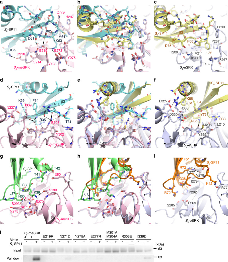
Fig. 2. Interface between S8-meSRK and S8-SP11.
a, d Close-up views of SP11-binding site 1 on S8-meSRK (pink) with S8-SP11 (cyan). g Close-up view of SP11-binding site 2 on S8-meSRK with another molecule of S8-SP11 (green). b, e, h Comparison of SP11-binding sites of eSRK between the S8-meSRK–S8-SP11 and S9-eSRK–S9-SP11 complexes. The eSRK molecules were superimposed using Cα atoms and show the same views as in a, d, and g, respectively. S9-eSRK is shown in silver, and S9-SP11 in yellow (interacting with SP11-binding site 1) and orange (site 2). c, f, i Close-up views of S9-eSRK–S9-SP11 complex in the same orientation as in (b), (e), and (h). a–i, Dotted lines represent hydrogen bonds. Water molecules are shown as small cyan spheres. j Pull-down of S8-meSRK-HLH mutants with biotin-S8-SP11 as in Fig. 1b.

