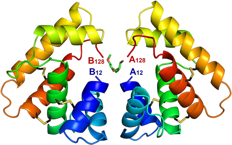
Figure 2.
Crystal structure of EposPBP3 with two molecules in the asymmetric unit. Each molecule is shown in ribbon mode and is rainbow coloured from blue (N-terminal) to red (C-terminal). The residue numbers of the N- and C-termini of each protomer (A,B) are indicated. The disulphide bridges are drawn in stick mode, with carbon atoms in black and sulphur atoms in yellow. PEG molecules are drawn in stick mode, with carbon atoms in green and oxygen atoms in red. Image drawn using PyMOL Molecular Graphics System, Version 2.0 (https://pymol.org/2/).

