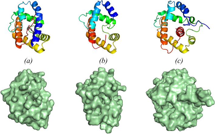
Figure 3.
Structure (top) and surface representation (bottom) of (a) the ligand-bound form (form B) of BmorPBP1 (1–137), bound to the natural pheromone bombykol (PDB 1DQE), (b) EposPBP3 (12–128) and (c) BmorPBP1 (1–142) in solution at pH 4.5 (form A) (PDB 1GM0). All structures are shown in ribbon mode and coloured from blue (N-terminus) to red (C-terminus). Bombykol is shown in sphere mode in (a), with carbon and oxygen atoms coloured dark grey and red, respectively. Image drawn using PyMOL Molecular Graphics System, Version 2.0 (https://pymol.org/2/).

