Fig. 1.

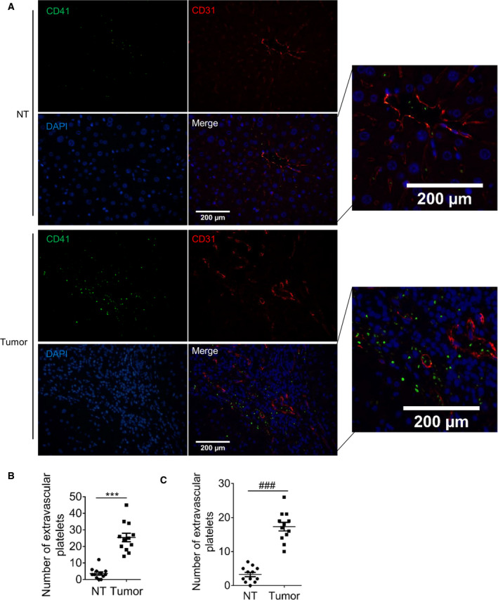
Platelets are present outside the blood vessels in HCC tissues. Human HCC tissues and adjacent nontumor (NT) liver tissues were stained with platelet‐specific marker CD41 (green), vascular endothelial molecule CD31 (red), and DAPI (blue). (A) The representative images (n = 4, random three fields per case). Bar, 200 μm. (B) The number of platelets. (C) Platelet number in C57 orthotopic tumor (n = 4, random three fields per case). Values are shown as dots and the mean ± SEM, ***P < 0.001, ### P < 0.001 (paired t‐test, 2‐tailed).
