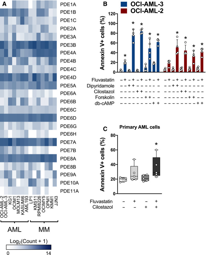
Fig. 3.

Compounds that increase cAMP levels phenocopy dipyridamole to potentiate statin‐induced apoptosis. (A) RNA expression of the different PDEs in a panel of human AML and MM cell lines. Data were mined from the CCLE database. (B) OCI‐AML‐2 and OCI‐AML‐3 cells were treated with fluvastatin (4 µm for OCI‐AML‐2 and 2 µm for OCI‐AML‐3) ± a PDE3 inhibitor (cilostazol; 20 µm), an adenylate cyclase activator (forskolin; 10 µm) or db‐cAMP (0.1 mm). After 48 h, cells were labeled with FITC‐conjugated Annexin V and apoptotic cells were quantified by flow cytometry. *P < 0.05 (one‐way ANOVA with Dunnett's multiple comparisons test, where the indicated groups were compared to the solvent controls group of that cell line). Data are represented as the mean + SD. (C) Primary AML cells were cultured in the presence of solvent controls, 5 µm fluvastatin, 20 µm cilostazol, or the combination. After 48 h, cells were labeled with FITC‐conjugated Annexin V and analyzed by flow cytometry. Data from four independent AML patient samples are represented as box plots with whiskers depicting the maximum and minimum values. *P < 0.05 (one‐way ANOVA with Dunnett's multiple comparisons test, where the indicated group was compared to the solvent controls group).
