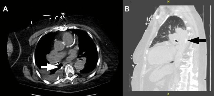
Figure 4.

(A) Axial slice of the computed tomography scan showing the radiopaque enteric tube positioned in the right bronchus and looping into the posterior pleural space (arrow). (B) Sagittal slice of the same computed tomography scan with a lung filter demonstrating the enteric tube lying in the posterior pleural space with surrounding lung field atelectasis and effusion (arrow).
