
Figure 1.
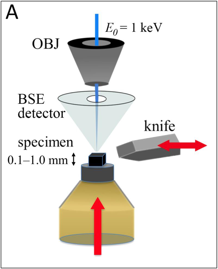
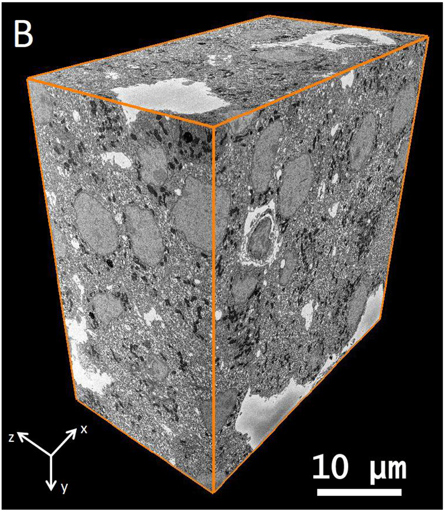
(A) A schematic of Serial Block-Face Scanning Electron Microscopy (SBF-SEM). After imaging with a back-scattered electron detector (BSD), a diamond knife cuts a thin slice from the face of the block. The newly exposed block face is then imaged, and the knife cuts again. This repetition of cutting and imaging allows one to build up a set of images, which represent the volume of the object imaged with the electron beam. (B) A part of wild type mouse islet 3D data set obtained by SBF-SEM. This data volume measures 42.6 μm × 43.5 μm × 23.9 μm in x y and z, respectively.