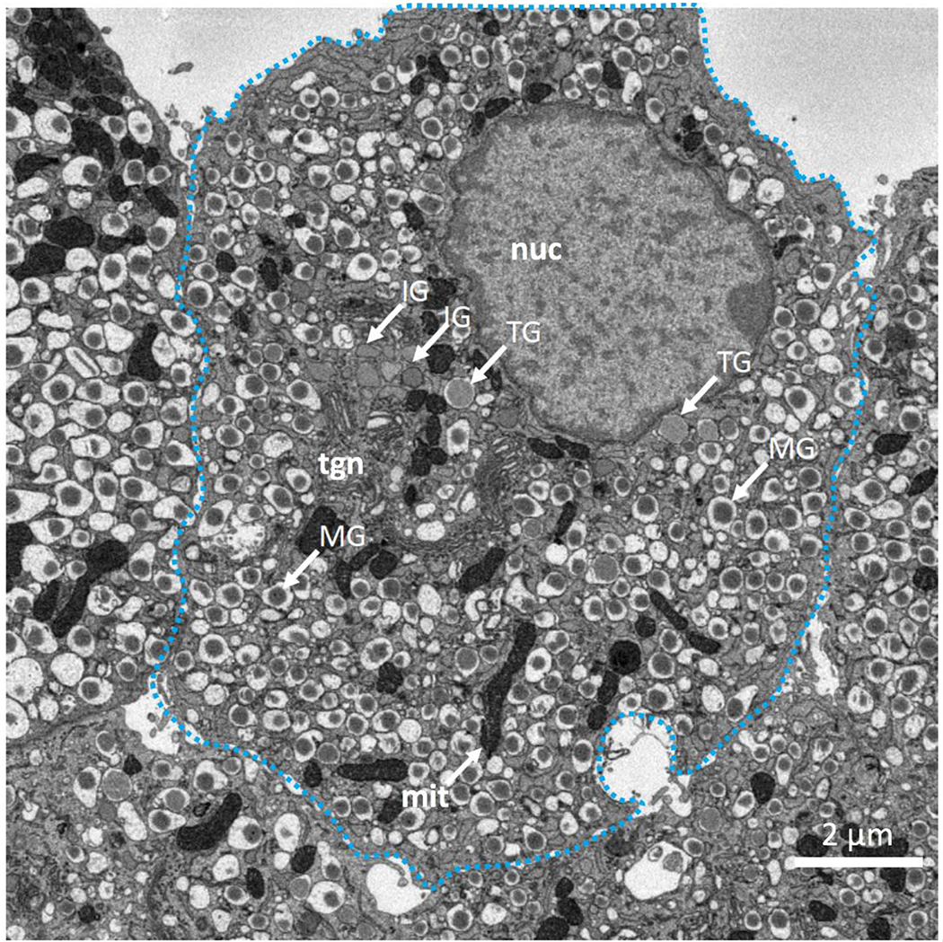
Figure 2.

(A) SBEM backscattered electron image of section through pancreatic β-cell indicating morphologies of mature secretory granules (MG), immature secretory granules (IG), and transforming secretory granules (TG); mature granules contain cores of condensed insulin with angular sides and large surrounding halos. Immature granules are situated mainly near the trans-Golgi network (tgn) and have no halos. Several of the immature granules are near nucleus (nuc), while others are interspersed with mitochondria (mit). The transforming granules form a distinct population with very narrow halos.
