
Figure 4.
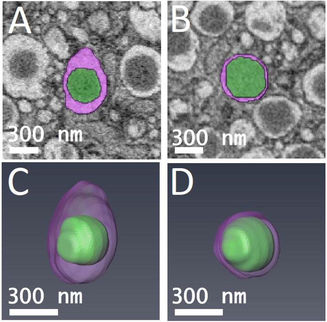
Segmented orthoslices of (A) mature β-cell secretory granule, and (B) transforming immature β-cell secretory granule. The granule cores are labeled in green, and the surrounding halos are labeled in purple. 3-D models of (C) mature β-cell secretory granule shown in (A), and (D) transforming immature β-cell secretory granule shown in (B). The ratio of core volume to total vesicle volume was measured for both granules.