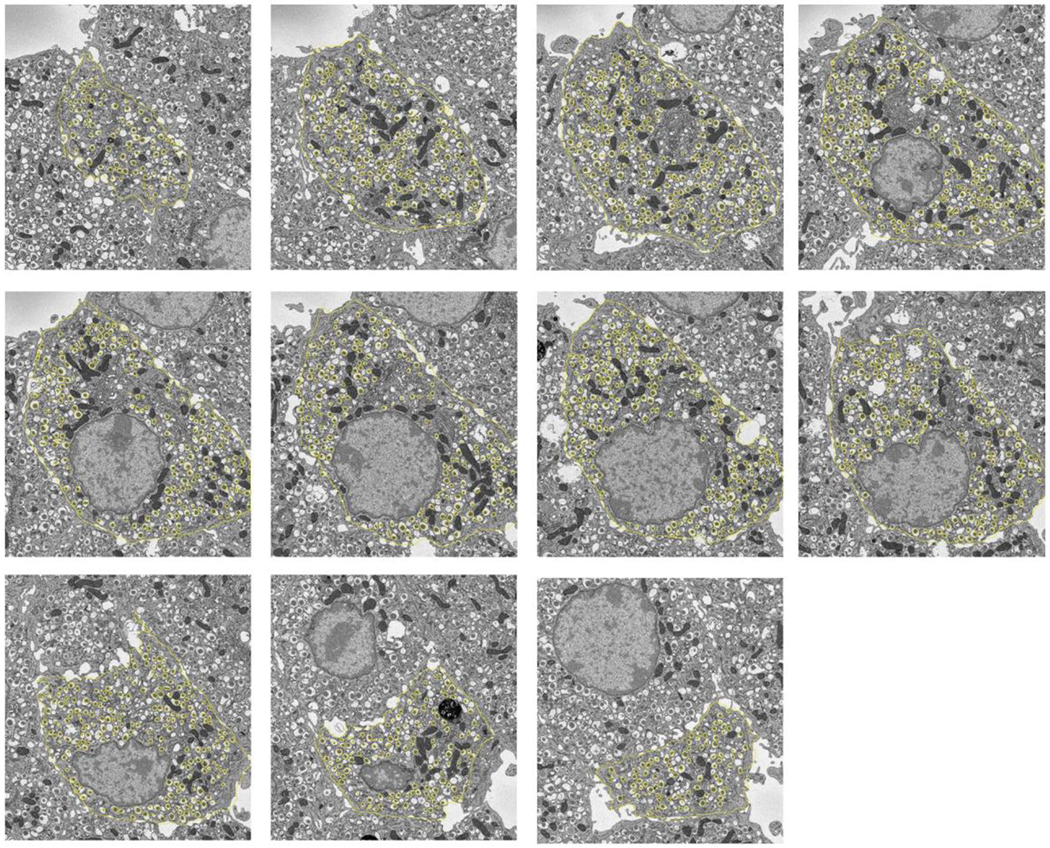
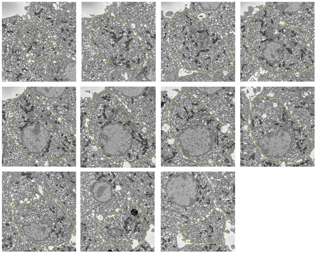
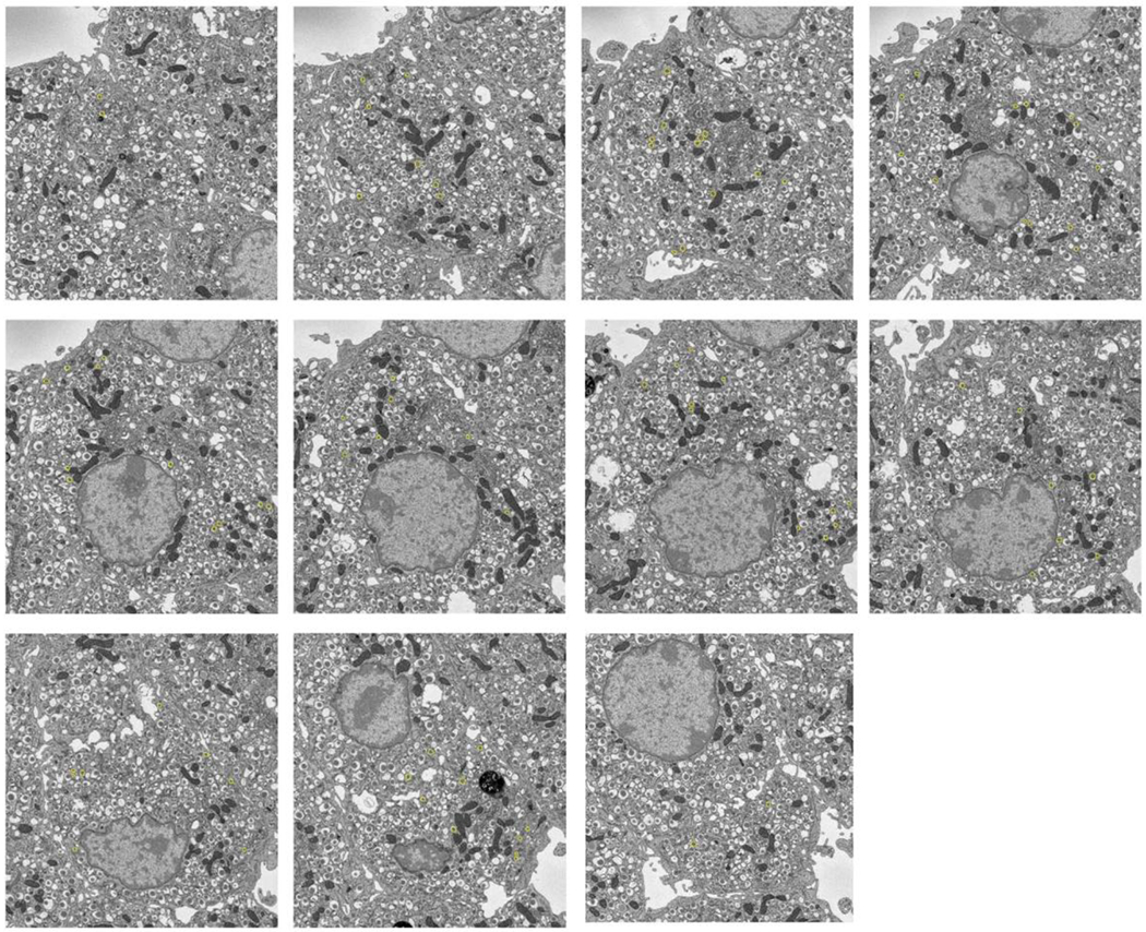
Figure. 6.


Segmentation of secretory granules in 11 slices spaced by 1 μm through a β-cell: (A) mature granules, defined by wide halos (typically ≳100 nm) and angular-shaped (crystalline) cores; (B) immature granules, most commonly located in vicinity of trans-Golgi network, and defined by absence of halos and amorphous cores; (C) transforming immature granules, defined by narrow halos (typically ≲30 nm). Cell membrane is outlined in (A) and (B). Full width of each image = 15 μm
